


Heal House Vitiligo Research 05 · Original Article
Can Wood's lamp vitiligo lesions be captured more clearly in photographs?
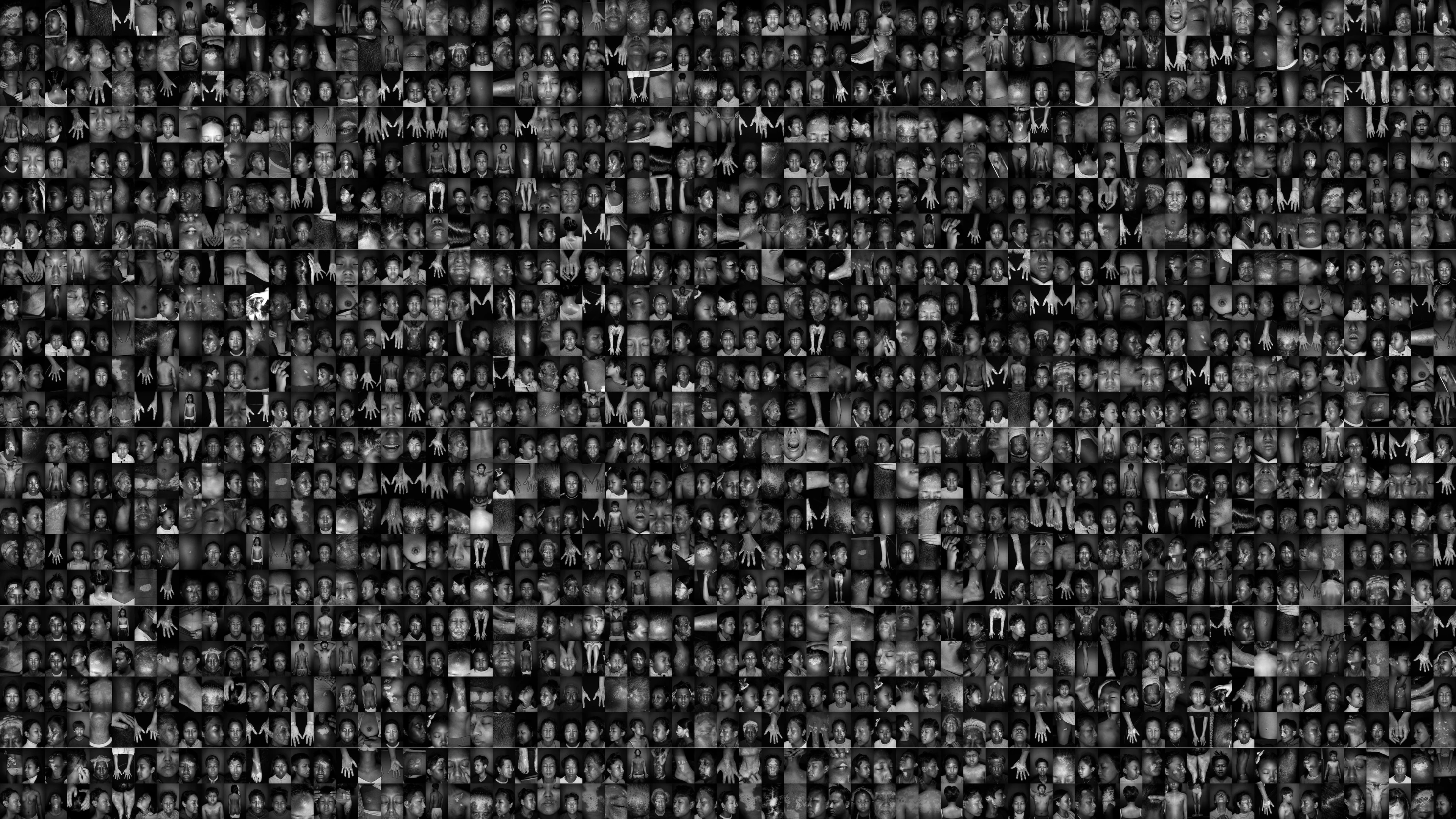
A grayscale photograph with high dynamic range taken under a Wood's lamp for better recognition of vitiligo lesions.
Last reviewed
In short
Converting photographs taken under a Wood's lamp to high-contrast black-and-white makes subtle vitiligo lesions and their borders much more distinct.
The Wood's lamp allows us to observe vitiligo well. However, it has always been regrettable that we cannot share the screen with you. Additionally, the bluish light characteristic of the Wood's lamp can appear unnatural. We wondered if there was a way to observe vitiligo more accurately and distinctly. This thought led to the development of ultraviolet photography. In conclusion, it allows us to record the current state of vitiligo and the progress of treatment more accurately and objectively, and we still find it very useful.
Q1 What is a high-contrast grayscale photo, and why does it show lesions more clearly?
We remove the color from photographs taken under a Wood's lamp and convert them to black and white. To this, we add a 'high-contrast (HDR)' process that emphasizes differences in brightness. Vitiligo lesions glow brighter than the surrounding skin under a Wood's lamp. However, in color photographs, this difference can easily be obscured by skin tone or lighting. By removing color information and retaining only brightness differences, the border between the lesion and normal skin becomes more clearly visible. Photographs processed in this way help to ensure that faint lesions are not missed. They are also useful for comparing treatment progress over time. Another major advantage is that it can be done with just photography and simple editing, without any special equipment.
Closing remarks
Although this is just a simple photography technique, it markedly improves the quality of medical records. In particular, by clearly recording the borders of faint lesions, we can much more accurately determine whether they have improved or spread at your next visit. It is also good for showing you the changes visually. I myself find this method very useful when tracking lesion changes in the clinic.